Non-traumatic Shoulder Instability
Mechanism of Onset:
The patient does not report any episode of falling onto the hand or hitting the shoulder forcefully.
Common Occupations and Gender:
This condition is more common in females from the mid-teens to their 40s.
• Frequently seen in individuals who use their upper limbs a lot, such as nursery school teachers, caregivers, or those who have played upper-limb-intensive sports like volleyball.
Main Symptoms:
- A sensation that the shoulder is about to slip out of place, an unpleasant feeling, and fear that prevents the patient from lifting the arm
- Severe pain at the moment of dislocation, sometimes accompanied by a popping or snapping sound
- Possible asymmetry in shoulder shape
- Inability to lift the arm due to pain
- A feeling of shoulder instability or looseness ("shoulder feels wobbly")
The typical presentation is shown in the video below.
Please watch the video. The patient is unable to raise the arm smoothly. They report that the shoulder feels unstable or "wobbly" when lifting the arm.
The humeral head is unstable. While raising the arm, the shoulder repeatedly subluxates and then reduces itself, resulting in this abnormal movement pattern.
What’s Happening in the Shoulder:
In patients with non-traumatic shoulder instability, there is often a mild labral tear where the ligament attaches to the glenoid—the socket that receives the head of the humerus.
Under normal conditions, the ligaments are kept taut to prevent the shoulder from slipping out of the socket (dislocating). However, when a labral tear occurs, the ligaments lose their tension.
As a result, the shoulder joint becomes unstable, leading to a sensation of looseness or "wobbliness." This instability can also be accompanied by pain.
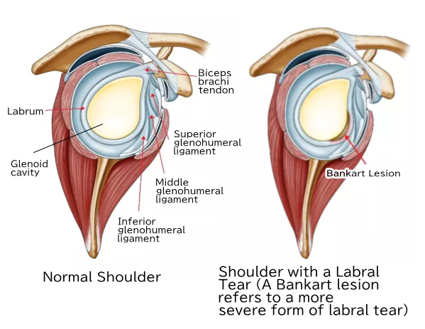
Note:
This illustration is taken from a depiction of recurrent shoulder dislocation. It has been used here to help clearly show the extent of a labral tear.
However, in actual cases, the tear is often only about 1 cm in size.
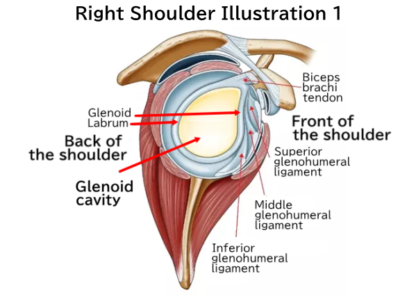
Additionally, labral tears can occur in various locations around the glenoid.
Therefore, we will now show the illustration again for reference.
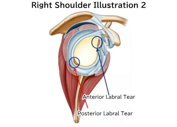
The area marked with a blue circle in the illustration above is a common site of injury.
In this area, labral tears can occur in a manner similar to a Bankart lesion, as shown in the illustration.
These may be anterior or posterior labral tears, depending on the location.
However, they are generally less severe than true Bankart lesions.
As a result, they typically do not cause repeated episodes of shoulder dislocation or subluxation.
Diagnosis: Physical Examination Findings
The diagnosis is made using an imaging test called contrast-enhanced MRI.
A saline solution mixed with contrast agent is injected into the shoulder joint, and MRI scanning is performed.
In the illustration below, the blue circle indicates where the contrast agent has collected — it leaks into the area of the injury.
Normally, the labrum is tightly attached to the glenoid.
If it is detached, fluid flows into the gap.
The contrast agent helps make these findings more visible on MRI.
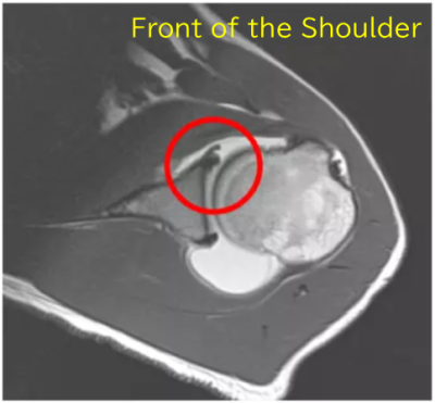
The red circle shows where the contrast agent has entered the area of injury.
The damaged area is small, and this finding is visible in only a few slices out of the many taken.
Treatment:
- For patients who cannot undergo surgery, a dislocation-prevention brace is prescribed and worn. They are instructed on the shoulder position that prevents dislocation. However, this cannot be considered an absolute treatment.
- Exercise therapy is prescribed to help patients recognize the shoulder position that prevents dislocation. In addition to strengthening the muscles around the shoulder, core strengthening and overall functional improvement are implemented.
Surgery:
Arthroscopic labral repair and stabilization procedures are performed.
The purpose of the surgery is to repair the labral tear.
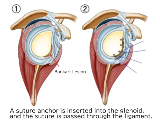
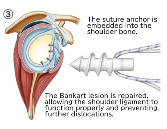
Note: The concept of the surgery is the same as for labral tears.
“A suture anchor (a screw with attached sutures) is inserted into the bone called the glenoid. The bone is attached to the labrum-ligament complex, and the sutures are then tied together to repair the tissue.”
Arthroscopic images will be shown.
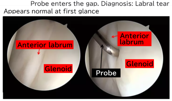
There is a metal rod called a probe.
It is used to examine the condition of tissues inside the joint, acting like a finger would.
Although the joint may appear normal at first glance, when the probe touches the labrum, its tip can enter the gap between the labrum and the glenoid.
This means that the labrum is not firmly attached to the glenoid, indicating the presence of a tear or injury.
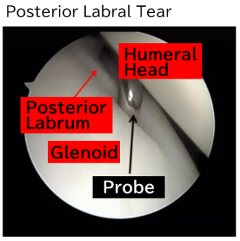
This is an arthroscopic image of a posterior labral tear.
Similar to the anterior labrum, the tip of the probe enters the gap between the posterior labrum and the glenoid.
This indicates the presence of a posterior labral tear.
Summary:
The diagnosis is very difficult.
In some cases, symptoms cannot be improved without surgery. However, even after surgery, recurrence can occur. Due to inherent ligament laxity, stress may cause the shoulder ligaments to loosen again.
It is recommended to have a thorough discussion with your attending physician about the benefits and risks.
Shoulder Diseases

We provide explanations for various shoulder conditions. Please use this as a general guide to determine which condition may apply to you.
- Common shoulder injuries by age group
- To those who neither have frozen shoulder nor rotator cuff tears
- Throwing Shoulder Disorder
- Rotator Cuff Tears and Rotator Cuff Injuries
- Recurrent Shoulder Dislocation
- Frozen Shoulder
- Shoulder Dislocation
- Acromioclavicular Joint Dislocation
- Chronic Acromioclavicular Joint Dislocation
- Frozen Shoulder
- Calcific Tendinitis of the Rotator Cuff
- Primary Degenerative Shoulder Arthritis
- Rotator Cuff Tear-related Degenerative Shoulder Arthritis
- Non-traumatic Shoulder Instability
- Biceps Tendon Injuries
- Surgical Trends for Throwing Shoulder in Baseball Players
